Nikon XT H 225ST CT Scanner (Large-Volume Micro-CT System)
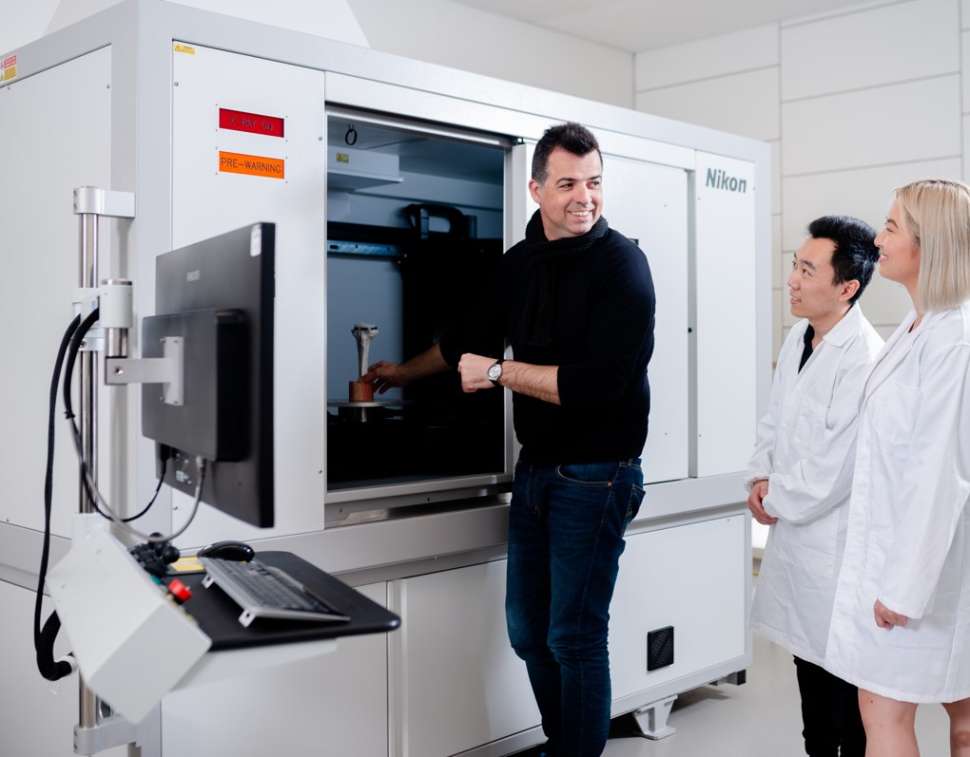
The Nikon XT H 225ST (Nikon Metrology, Tring, UK) is a large volume cabinet Computed Tomography (CT) system enabling the 3D-scanning of large and heavy samples for research and industrial applications. This includes whole machine parts, excised human and animal limbs/segments, prosthetic devices, batteries, plants, fossils, vertebrates and 3D composite materials. This also allows experimental testing rigs, such as mechanical stages (listed below) or environmental chambers, to be placed inside the scanner (in situ testing), for testing samples while scanning, to study the structure-function relationships.
Contact
Instrument Leader: A/Prof Egon Perilli
Micro-CT Facility Manager: Dr Sophie Rapagna

The X-ray source has a maximum power of 225W and maximum voltage of 225 kVp, allowing for the X-ray penetration of thick and dense materials. The system can scan samples up to 240 mm in diameter and 350 mm in height within the field of view (two stacked scans). The sample weight can be up to 50kg. The “field of view:pixel size” ratio is 4000:1, with resolution from 5 µm to 60 µm (or 240 µm, depending on the binning). For example, an object sized 100 mm x 100 mm (diameter x height) can be entirely scanned at 25 µm pixel size and a 200 mm x 200 mm object at 50 µm pixel size. Helical scanning modality is also available, allowing long samples (up to 220 mm length) to be scanned without stitching artefacts, such as an 80 mm diameter x 196 mm height object at 20µm pixel size.
The reconstructed cross-section images come as stacks of grey-level bitmap or .tiff files, displaying the internal specimen density and microarchitecture. These can be used for visualization of the specimen internal details in any plane, creation of 3D models and calculation of morphological parameters.
We have two mechanical testing stages available that can be used with the micro-CT system. These allow tensile and compressive loading of the specimen while scanning: an in-house-built stage (20kN load cell) and a customized stage (5kN load cell, Deben CT5000N, Deben, UK).
Digital volume correlation software (available software: DaVis, LaVision) can be used, to quantify displacements and strains from specimens scanned in loaded versus the unloaded conditions.
The system can scan samples up to 240 mm in diameter and 350 mm in height within the field of view (two stacked scans). The sample weight can be up to 50kg. Examples of applications include, but are not limited to:
- whole machine parts
- excised human and animal limbs/segments
- prosthetic devices
- batteries
- plants
- fossils
- vertebrates
- 3D composite materials.
Micro-CT in the news
Application examples


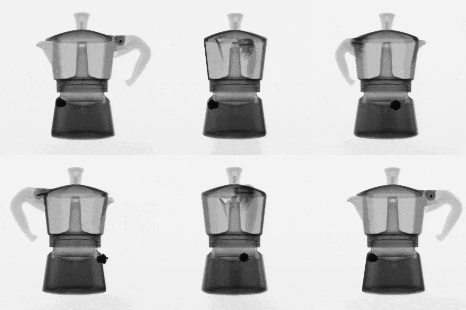
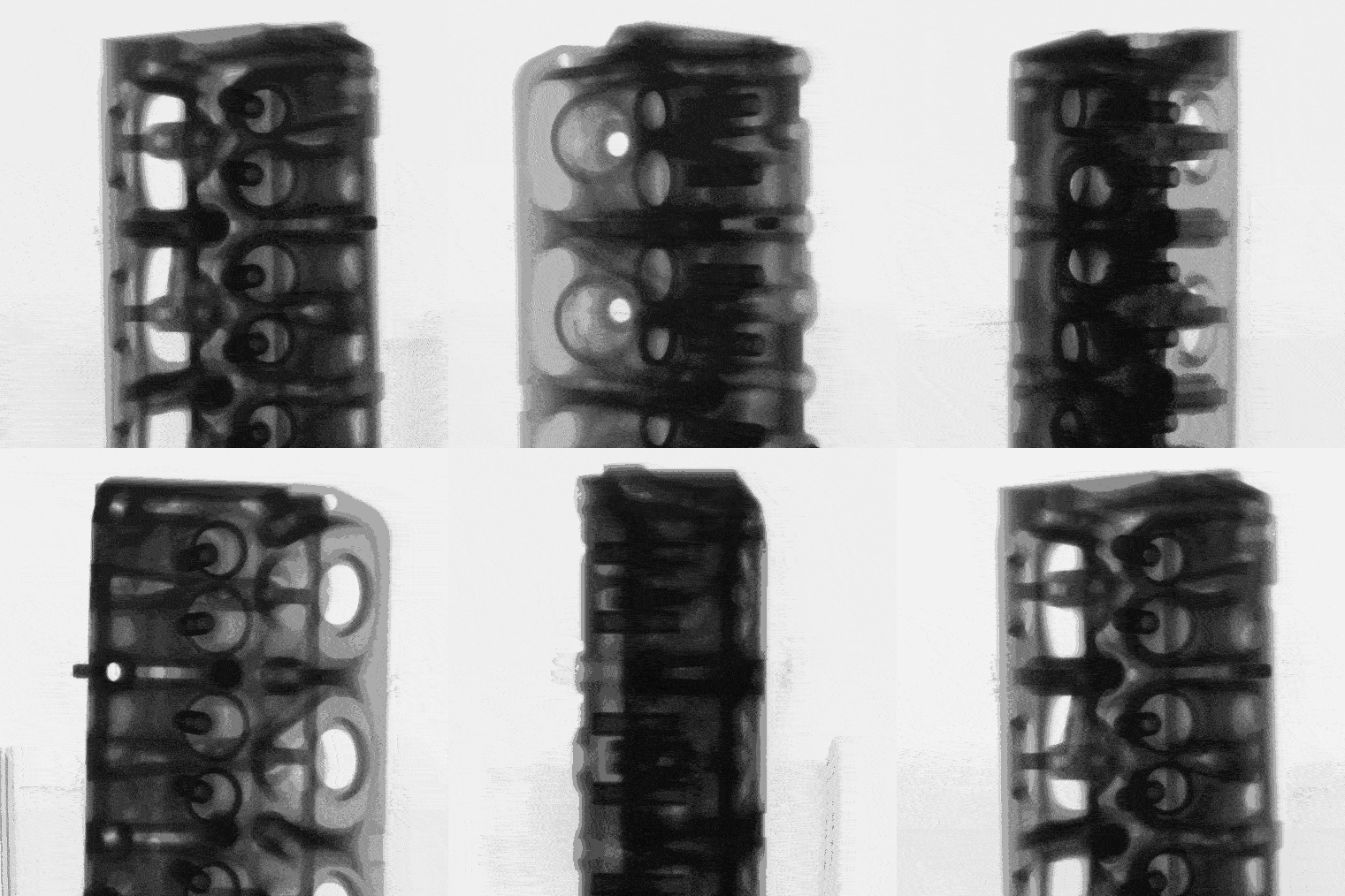
X-Ray projections over 360° of an aluminum moka pot (left) and cylinder head (right)


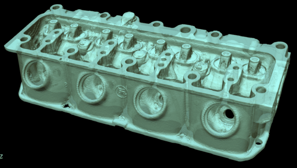
Micro-CT 3D rendering of a human proximal femur (24µm/pixel; left) and an aluminum cylinder head (121µm/pixel; right)
Our equipment is funded by:


![]()
Sturt Rd, Bedford Park
South Australia 5042
South Australia | Northern Territory
Global | Online
CRICOS Provider: 00114A TEQSA Provider ID: PRV12097 TEQSA category: Australian University


