Atomic Force Microscopy
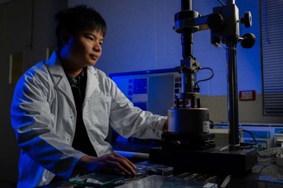
Atomic Force Microscopy (AFM) is used to gain topographic information on a sample. The AFM operates by scanning a sharp silicon tip across the sample surface effectively tracing it. The AFM then combines a series of traced lines or cross-sections to produce a 3D image of the sample. The AFM can also operate in numerous imaging modes which allow the measurement of surface adhesion, stiffness and sample conductivity on the nanoscale. The types of materials analysed by AFM in the FMMA include, for example, 2D materials, nanotubes, nanoparticles, polymers, metals, and semiconductors.
Contact
Instrument Leader: Dr Chris Gibson

FMMA has two Bruker Multimode AFMs and one Bruker Dimension AFM. Capabilities are as follows
- Lateral resolution 5-10 nm (dependant on AFM tip diameter, vertical resolution less than 1 nm.
- Contact mode
- Tapping mode
- Force spectroscopy
- Peakforce tapping mode
- Peakforce TUNA mode – conductance imaging
Application examples

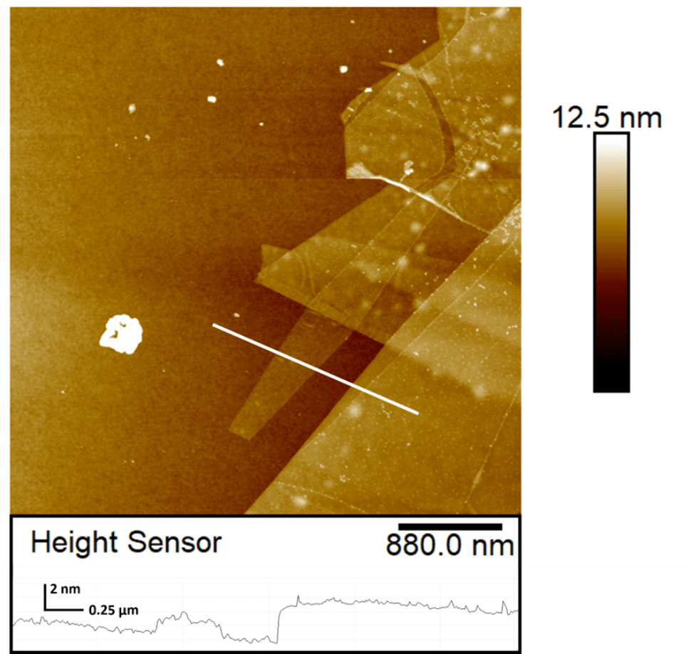
An AFM topography image of a flake of graphene. This flake contained single layer and multi-layer graphene ranging in thickness from 0.34 to 1.7 nm and demonstrates the high-resolution capability of these types of microscopes. The cross section underneath the AFM height image corresponds to position of the while line and intersects a flake of single layer graphene.
Our equipment is funded by:


![]()
Sturt Rd, Bedford Park
South Australia 5042
South Australia | Northern Territory
Global | Online
CRICOS Provider: 00114A TEQSA Provider ID: PRV12097 TEQSA category: Australian University


