Industry solutions
Flinders Microscopy and Microanalysis offers a range of characterisation services to businesses, from small start-ups to multinational companies.
We are a multidisciplinary team.
We are experts in biology, chemistry, physics, nanotechnology, geology, materials science and engineering. If you have a research or development project you want to pursue, we want to help you foster innovation. We will work with you to customise our testing and consultancy services to your needs. You can draw on our research and technical expertise, access our microscopy facilities and equipment, and even access a whole new range of government and research grants.
Let us help you develop new products or processes.
We can improve your existing products and processes—or help you create new ones. We are able to provide high resolution imaging and analysis, resulting in firm outcomes and recommendations. Our past projects have looked at the surface properties of materials to prevent corrosion and corrected defects in 3D printing technology. Typically we work with polymers, particle coatings and films, alloys and metals, plastics, and many others. We find and resolve defects and investigate ways to improve what's already working.
Get in touch to find out how we can help you.
Working with industry and developing new techniques

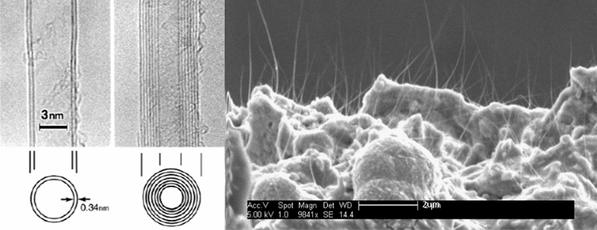
Micro-X Ltd designs, develops, and manufactures a range of innovative, ultra-lightweight, mobile x-ray imaging systems for medical and security applications. The competitive advantage of these miniaturised products stems from a new technology of electronic x-ray tube using carbon nano-tube (CNT) field emission devices, enabling the miniaturisation of a number of x-ray applications relevant to large global markets.
Demands for high throughput and resolution medical and inspection techniques such as radiotherapy, computed tomography and tomosynthesis, security inspection and high throughput manufacturing has resulted in a considerable level of interest in x-ray sources capable of meeting these requirements.
Micro-X has a contract from the Australian Department of Defence to develop and demonstrate the technology of an ultra-lightweight, digital mobile x-ray which is optimised for use in military deployed medical facilities. The product also has potential applications in humanitarian aid and disaster relief. The third product in Micro-X’s development pipeline is a mobile backscatter imaging device in the form of a miniature x-ray system allowing for stand-off imaging of improvised explosive devices.
Collaboration with the Flinders Institute for Nanoscale Science & Technology, with help from a business innovation grant from the Department of Innovation Science and Technology, has played an important role in accelerating the development of core technologies needed in Micro-X’s next-generation products. Tapping into Flinders’ vast expertise and the equipment through Flinders Microscopy and Microanalysis has provided Micro-X with a better understanding of their systems and refined advanced manufacturing processes. Micro-X has a goal to develop, source and manufacture all components from Australia and create jobs in South Australia.

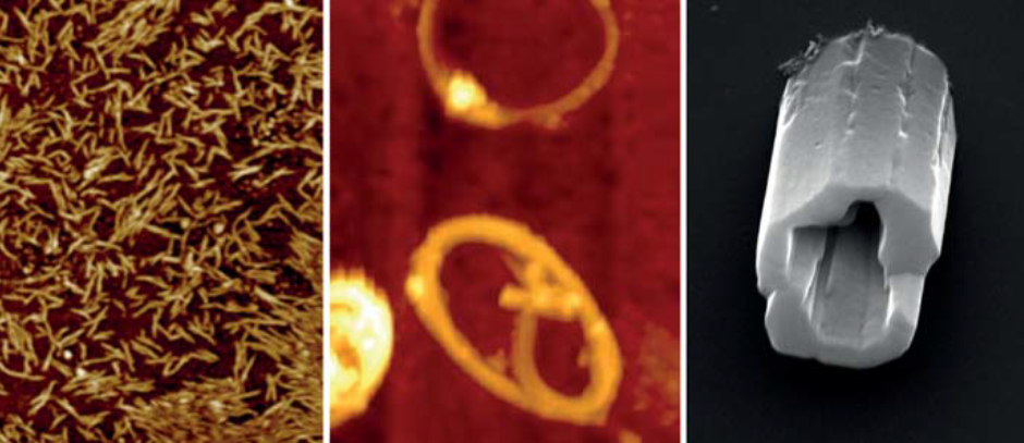
Atomic force microscopy shows SWCNT (left) and SWCNT rings (centre). Pictured right: SEM image of buckyball tubules.
Professor Colin Raston grabbed world headlines in 2015 when he won the Ig Nobel Prize for the development of a ‘Vortex Fluidic Device’ (VFD) that can unboil an egg.
The VFD is a seriously useful invention. It can be used to refold misshapen proteins, produce biodiesel from waste oils, modify the characteristics of wine and facilitate a range of different chemical transformations. Professor Raston’s group is using the VFD to create carbon nanostructures that could increase the efficiency of solar cells and improve polymer composites, sensing devices, electronics and drug delivery.
Dr Kasturi Vimalanathan is working with Professor Raston on these carbon nanostructures, which are made of graphene—single layers of graphite. Single-walled carbon nanotubes (SWCNT) can be cut into specific lengths by using a pulsed laser with various solvents and altered shear forces in the VFD. The VFD can also bend SWCNTs into rings without reactive chemicals or stabilising surfactants. The diameter of the nanorings is controllable—either from 100 to 200 nm or 300 to 700 nm—and production can be readily scaled up.
Dr Vimalanathan has also used the VFD to assemble nanoscale carbon spheres, commonly known as buckyballs (C60), into crystalline nanotubules without stabilising agents and without trapping solvent molecules during crystallisation. The VFD efficiently controls the assembly and can form micrometre-length nanotubules with a hollow diameter of 100-400nm.
During these manipulations, atomic force microscopy and scanning electron microscopy in the Flinders Microscopy and Microanalysis facilities were used to visualise and measure the nanoscale products.

Graphene consists of flat layers of carbon and has emerged as a material with a vast variety of applications. The electronic, optical and mechanical properties of graphene are strongly influenced by the number of layers present in a sample.
As a result, the dimensional characterisation of graphene films is crucial, especially in terms of the thickness and number of layers. Atomic force microscopy (AFM) is often used to determine the thickness of single layer graphene films but a wide range of values from 0.4 to 1.7 nanometres have been reported in the literature.
The major issue with imaging one-atom thick materials is that there is rarely a perfect contact between the substrate and sample. This imperfect contact can be further exacerbated by the presence of a single layer of water molecules, often present on all surfaces under standard conditions. This issue is most commonly observed when imaging with an atomic force microscope (AFM), which directly images a sample in three dimensions using an atomically sharp tip.
Researchers at Flinders University, led by Dr Cameron Shearer and Dr Christopher Gibson, have optimised an AFM technique called PeakForce Tapping AFM to accurately measure graphene by imaging with high force. At low applied force, the measured height is equivalent to the sum of the graphene layer thickness plus that of the intervening water. As the force applied to the graphene by the AFM tip increases, the liquid is gradually pushed out of the way until finally the graphene makes direct contact with the underlying substrate and a much more accurate value is measured. This work was published in the journal, Nanotechnology.

The expertise of Flinders Micrsocopy and Microanalysis, along with researchers from other Micrsocopy Australia facilities and corporate partner, FEI, came together to develop an interactive virtual microscopy experience. Whether you want to view projections on a vaccine nanopatch, surface details of a dinosaur egg or air bubble distribution in frozen ice cream, you can learn to use a scanning electron microscope faster with MyScope. This innovation in training for advanced research offers virtual instruments, step-by-step instructions and expert knowledge. Additional modules covering transmission electron microscopy, x-ray diffraction, scanning probe/atomic force microscopy, light/confocal microscopy, and microanalysis are also available, along with a free Outreach platform designed for ages eight and up.
There is growing demand for high-end education in science and technology across Australia and internationally. Freely available to all, MyScope provides researchers and students with a flexible, individual learning path that can be integrated with traditional learning environments.
'We hope that it will help get more young students excited about entering into science, technology, engineering and math careers,' said John Williams, FEI.
Developed in consultation with science educators in Australia and North America, MyScope Outreach enables discovery at the micro and nanometre scale. Interactive learning is enriched with audio, animations and hands-on activities. Please help us spread the word to promote this innovative STEM resource and bring the thrill of microscopy to Australia’s future scientists.
What we provide
- Expert recommendations
- Prompt and professional service
- Advanced microscopy techniques
- A process and outcomes customised to your needs
- Creative thinking and problem solving
- Training provided by leading instrumentation experts
- Links with government-funded facilities, such as Microscopy Australia and the Australian National Fabrication Facility (ANFF)
What you can expect
- Sample preparation
- Use of advanced microscopy techniques
- Optical and biomedical imaging
- Materials characterisation
- Image processing
- 3D modelling
- Data analysis
The techniques we use
- Metastable induced electron spectroscopy
- Scanning auger nanoprobe
- UPS
- IPES
- XPS
- Atomic force microscopy
- Tip enhanced raman spectroscopy
- Confocal raman microscopy
- Scanning electron microscopy
- X-ray photoelectron spectroscopy
- Neutral impact collision ion scattering spectroscopy
- Confocal fluorescence imaging
Our industry partners work in
- Defence
- Food and agriculture
- Manufacturing
- Maritime
- Technology
Our equipment is funded by:


![]()
Sturt Rd, Bedford Park
South Australia 5042
South Australia | Northern Territory
Global | Online
CRICOS Provider: 00114A TEQSA Provider ID: PRV12097 TEQSA category: Australian University


