Scientific image competition - past winners
Researchers from our institute capture hundreds of images every year as they explore problems and processes on a nanoscale. These amazing images are rarely shared outside of the research context.
Our Scientific Image Competition is a chance to celebrate the unique images produced, as well as the diverse research behind them.
Winner - Scientific Image of the Year
In late 2019 an expert judging panel including Monique Russell from Blend Creative Design Agency, and Emeritus Professor Ian Gibbins voted for the Scientific Image of the Year.
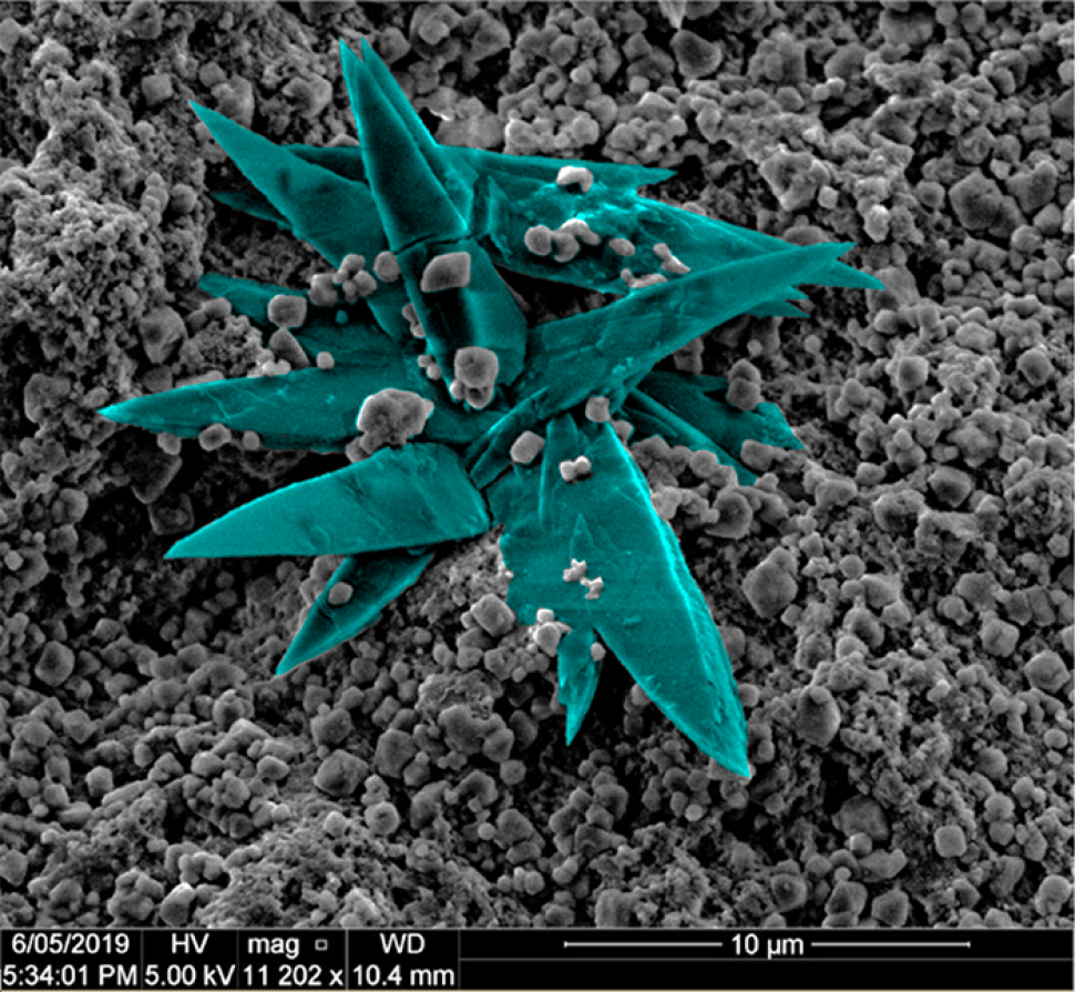
Zhen He, a PhD candidate supervised by Professor Sarah Harmer, took home the honour and was awarded a gift voucher valued at $200. His winning image, Formation of covellite by Zhen He will be used for the Institute for Nanoscale Science and Technology annual report cover.
Winner - People’s Choice Award
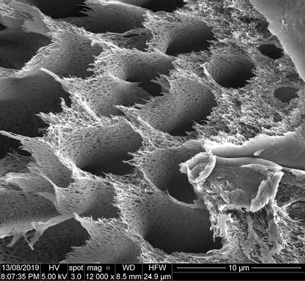
For the first time in the competition’s history, a People’s Choice Competition/Award was announced. With over 200 voters participating, it was Aghil Igder’s, image Nano Beehives, a membrane fabricated in the Vortex Fluidic Device electron microscope, that was the stand out winner.
Aghil, a PhD candidate supervised by Professor Colin Raston, will enjoy lunch at Flinders multi award-winning café, Alere.
1. A silver STEM
Samantha Pandelus
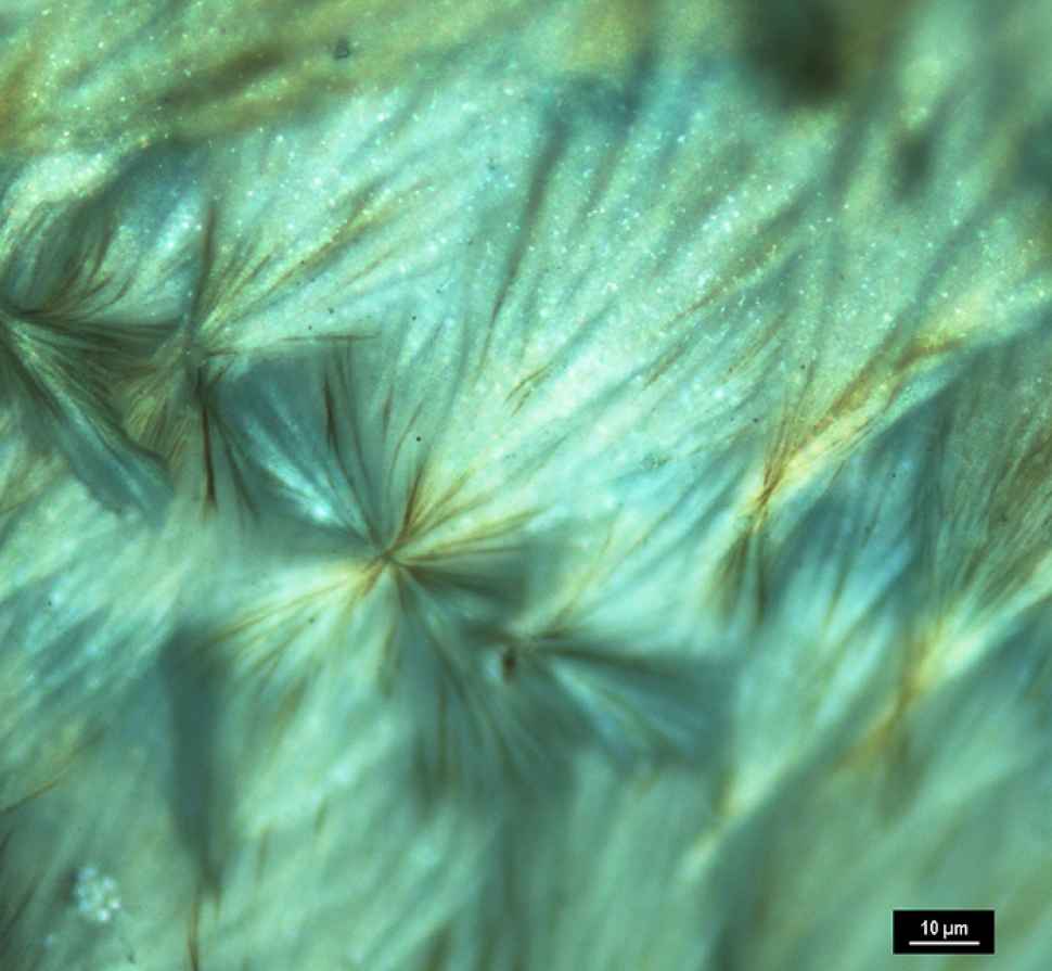
A stem cross section from a native Australian ‘hop bush’ plant coated with a silver photographic gel.
2. Alginate flower
Javad Tavakoli
One-step fabrication of alginate hydrogel microneedles using the Vortex Fluidic Device where a sodium alginate solution is injected in a calcium chloride solution in an angled rapidly rotating tube. For the first time, we found a significant change in the surface morphology of the alginate hydrogel films using different feed inlet diameter and rotation speeds. The proposed onestep facile fabrication of alginate films with different surface morphologies, has tangible potential for applications in the biomedical field including cell- surface studies and sustainable drug delivery.
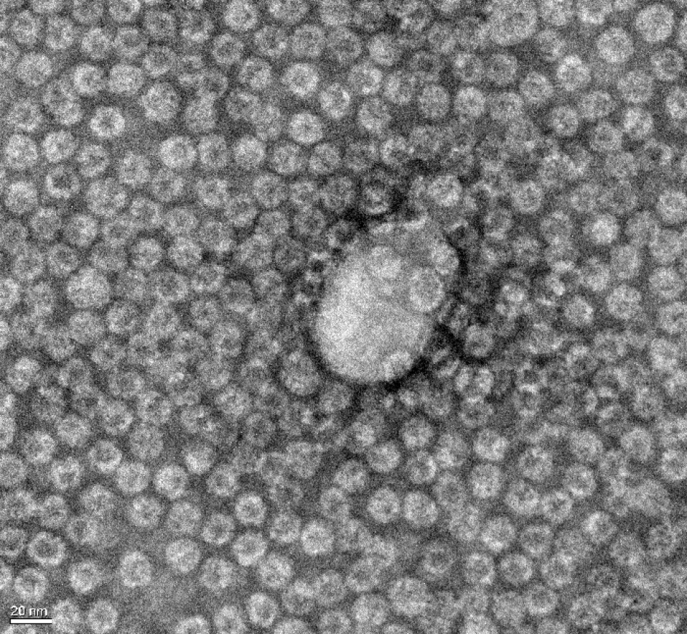
3. Biomolecular based smart nanocarriers for drug delivery
Gaurav Singhai
Successful assembly of virus protein over DNA nanostructures (two biomolecules) in the centre. The excess of empty small spherical virus-like particles with homogenous size confirms the assembly. These assembled particles have direct application as smart carriers in the field of nanomedicine.
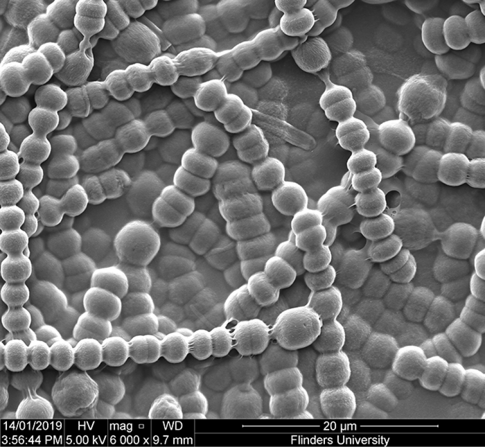
4. Cyanobacteria
Karuna Skipper
A film of cyanobacteria.
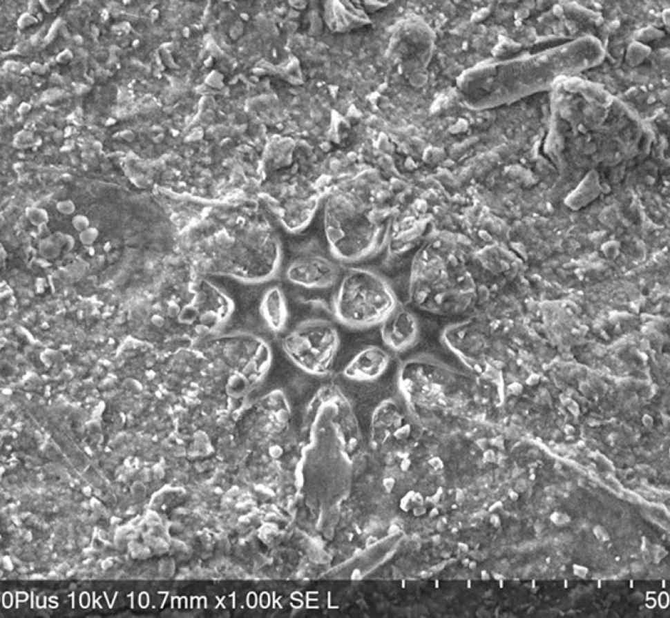
5. Diatoms in Flinders Lake
Danielle Saunders
The image is of a cluster of diatoms from the Flinders University lake. I conducted a preliminary with Dr. Martin Cole from NewSpec, to test TM4000 series electron microscope.

6. Formation of covellite (CuS) by replacing chalcopyrite (CuFeS2)
Zhen He
Copper sulfide (CuS) minerals formed by replacing chalcopyrite (CuFeS2).
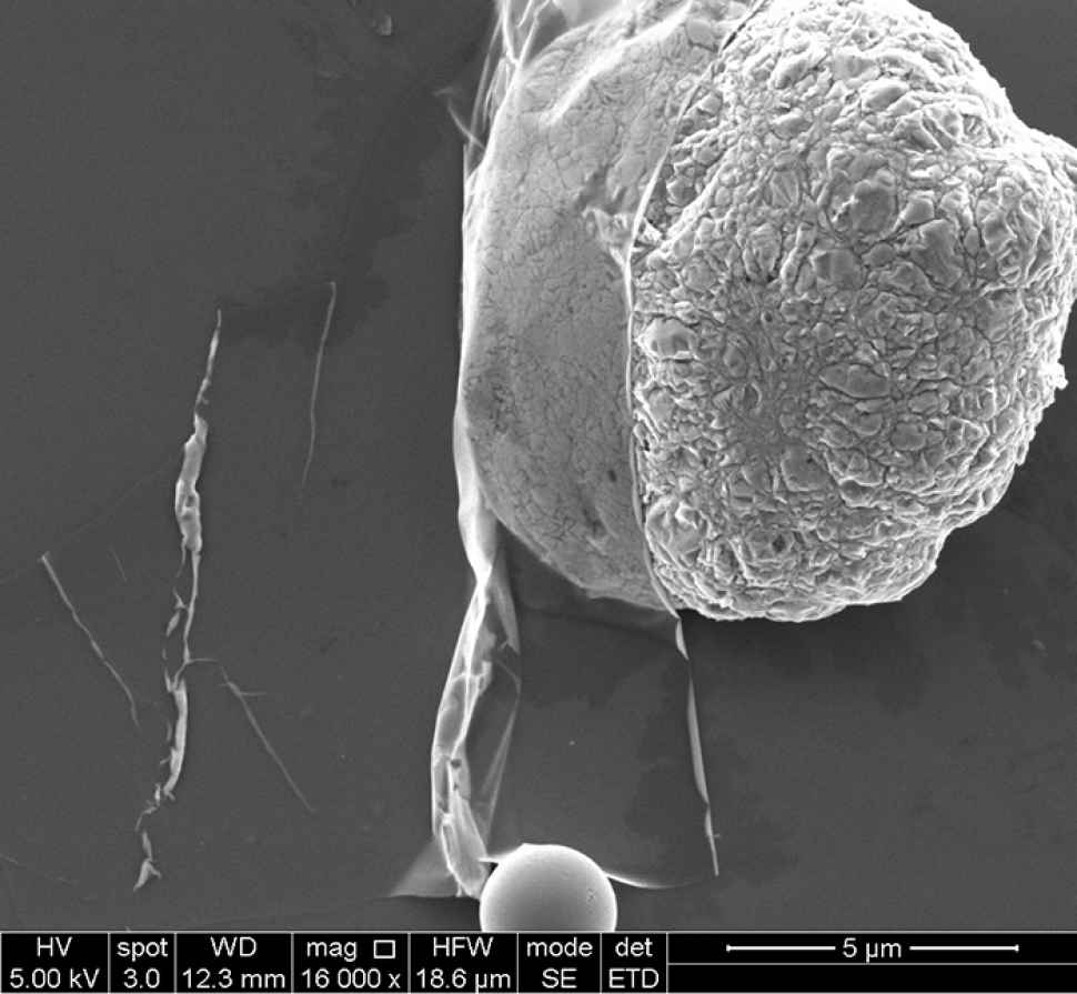
7. Gallenene sheet
Kasturi Vimalanathan
Transparent gallenene sheet sitting on a gallium sphere.
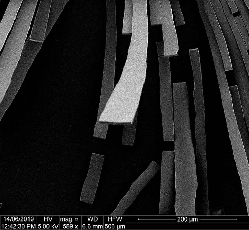
8. Inception
Stefan Martino
The individual “planks” are formed from silica nanoparticles clumped together that lifted off the surface as they dried.
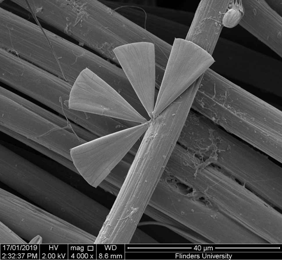
9. I'm your smallest fan
Karuna Skipper
A fan shaped diatom growing on carbon cloth.
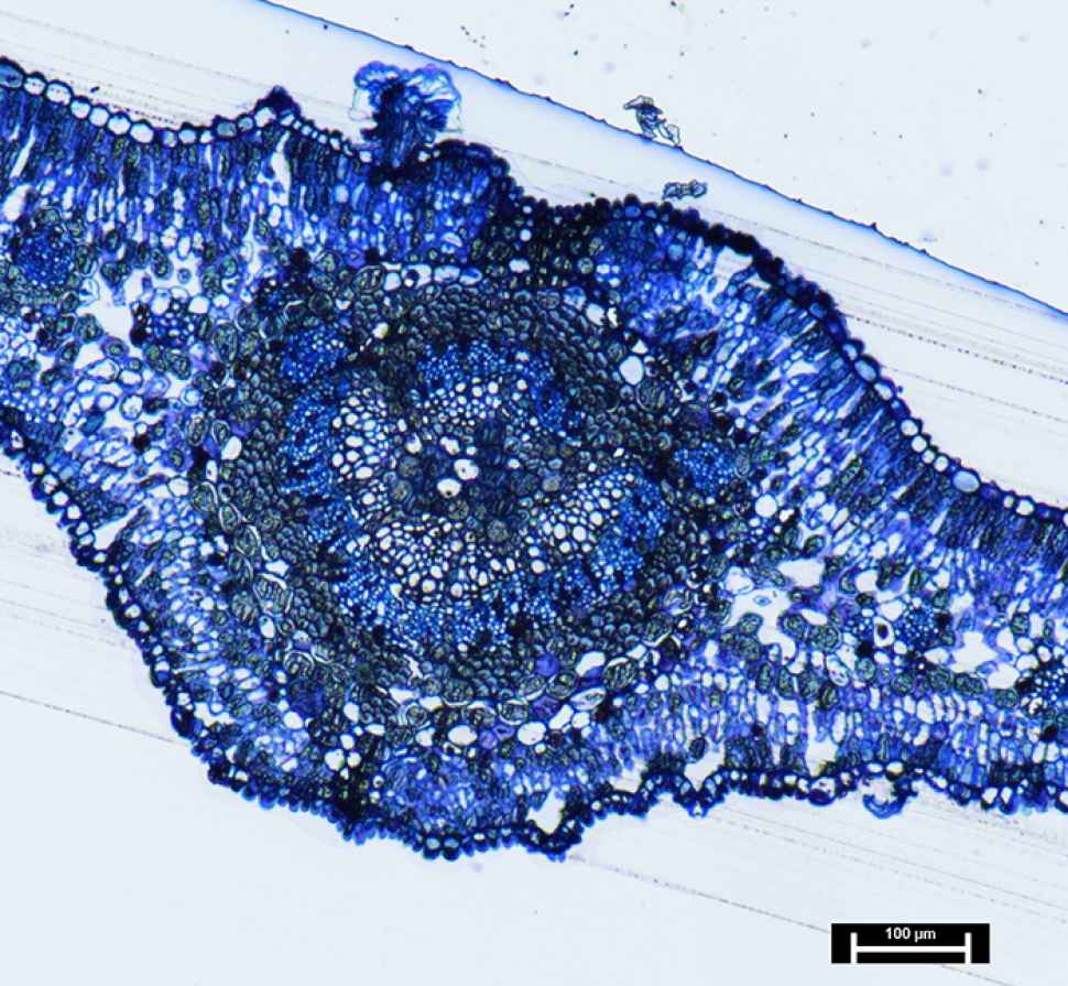
10. Leaf cross section
Samantha Pandelus
A cross section of a leaf of the native Australian ‘Hop Bush’ plant. The leaf was embedded with resin, cut with a glass knife and stained blue to show the cell structure of the leaf.
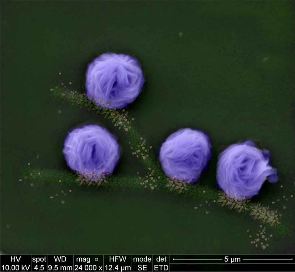
11. Nanoflowers
Xuan Luo
A hierarchical flower-like structure with hundreds of nanopetals is developed by mixing copper sulphate with a protein (bovine serum albumin or BSA) in a vortex fluidic device (VFD). This single step process significantly reduces the conventional processing time from 3 days into half an hour, which is ready to be used for enzymatic reactions and diagnostic sensors

12. Nano beehives
Aghil Igder
VFD mediated Polysulfone (PSF) filtration Membrane.
A cross section SEM imaging of a Polysulfone (PSF) filtration membrane fabricated from a phase-inversion process after micromixing the polymer solution in the Vortex Fluidic Device.
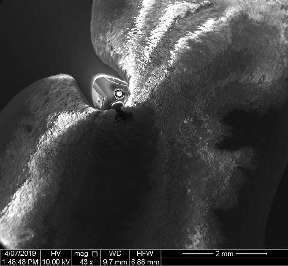
13. Peek-a-boo!
Steve Trewartha
A reflection on ceramic adhesive caused an image of the electron gun in the SEM to form, appearing to peak around the warped image of the ceramic surface.
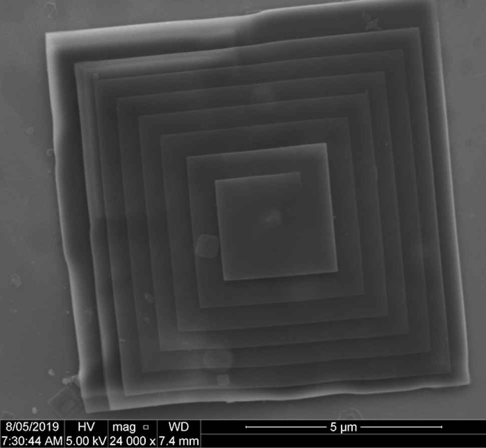
14. Pyramid
Ahmed Al-Antaki
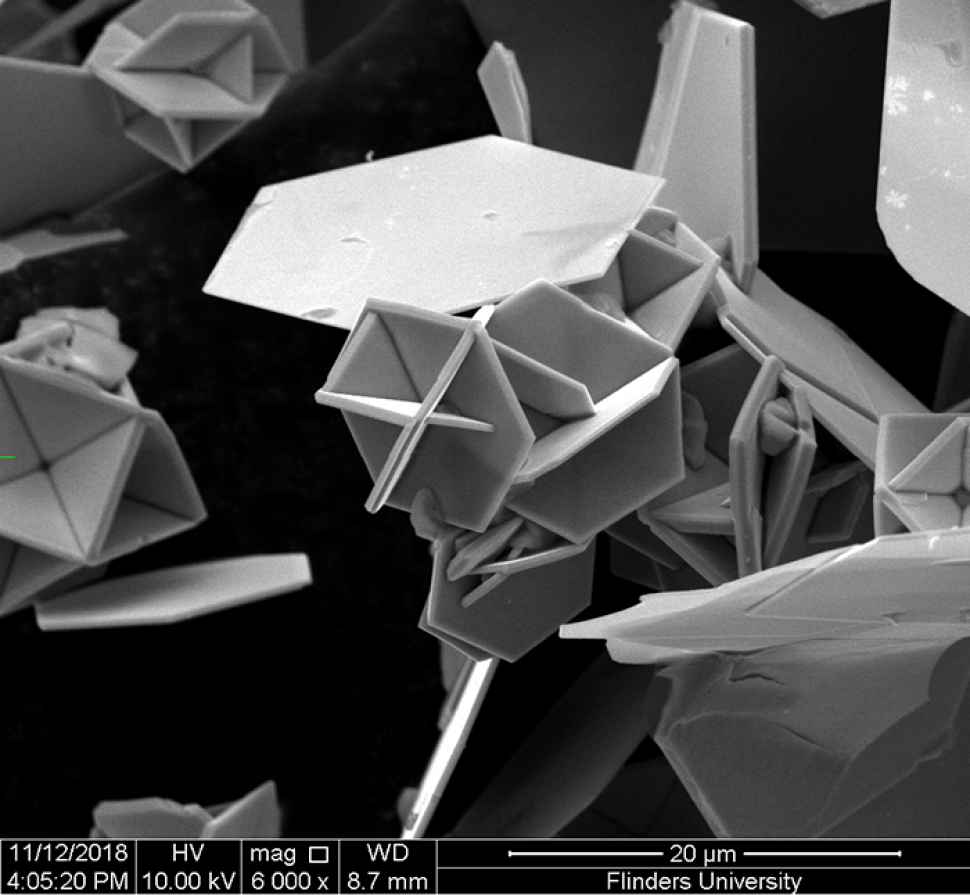
The shear stress in the dynamic thin film of the VFD has the ability of fabricated p-Phosphonato calix[4]arenes.
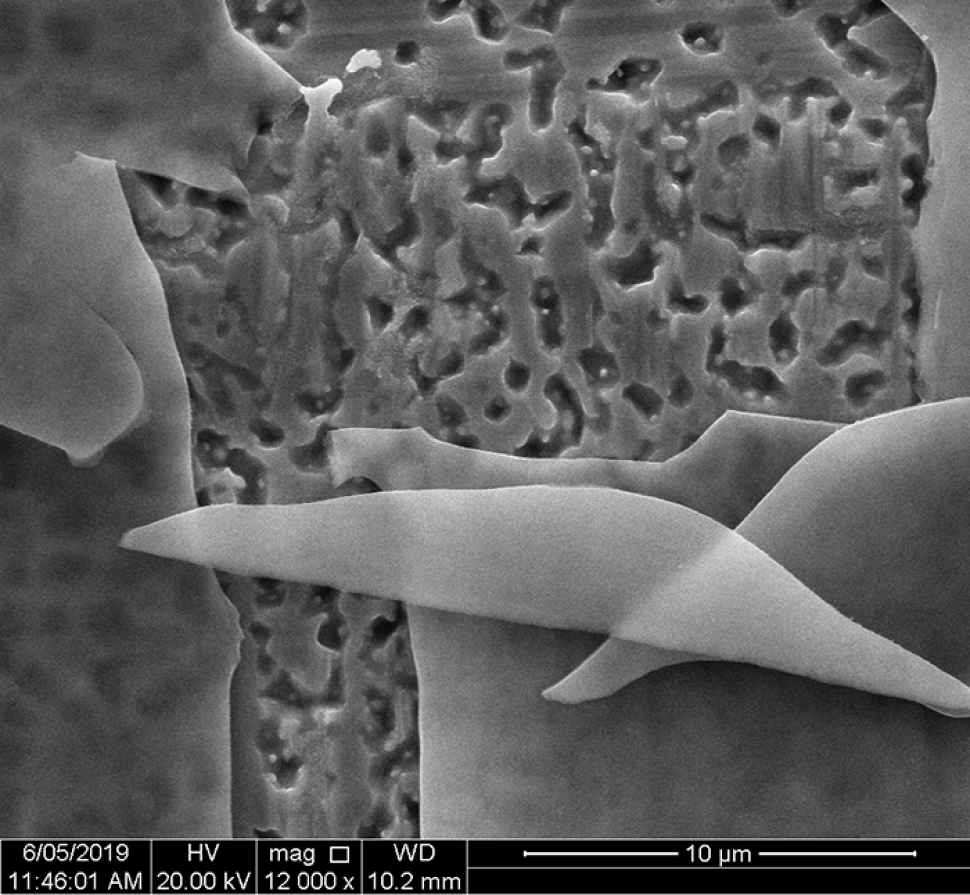
15. Shattered
Susanne Sahlos
Scanning electron microscope image of a shattered oxide layer that formed on the surface of stainless steel during a high temperature heat treatment.

16. Simulation of high-output sliding-mode triboelectric nanogenerators
Mohammad Khorsand
Recently triboelectric nanogenerators (TENGs) have been introduced to be a sustainable candidate in energy harvesting applications. With the help of artificial intelligence, we numerically predicted the outputs of TENGs, and presented a high-powered and lightweight generation of TENGs.
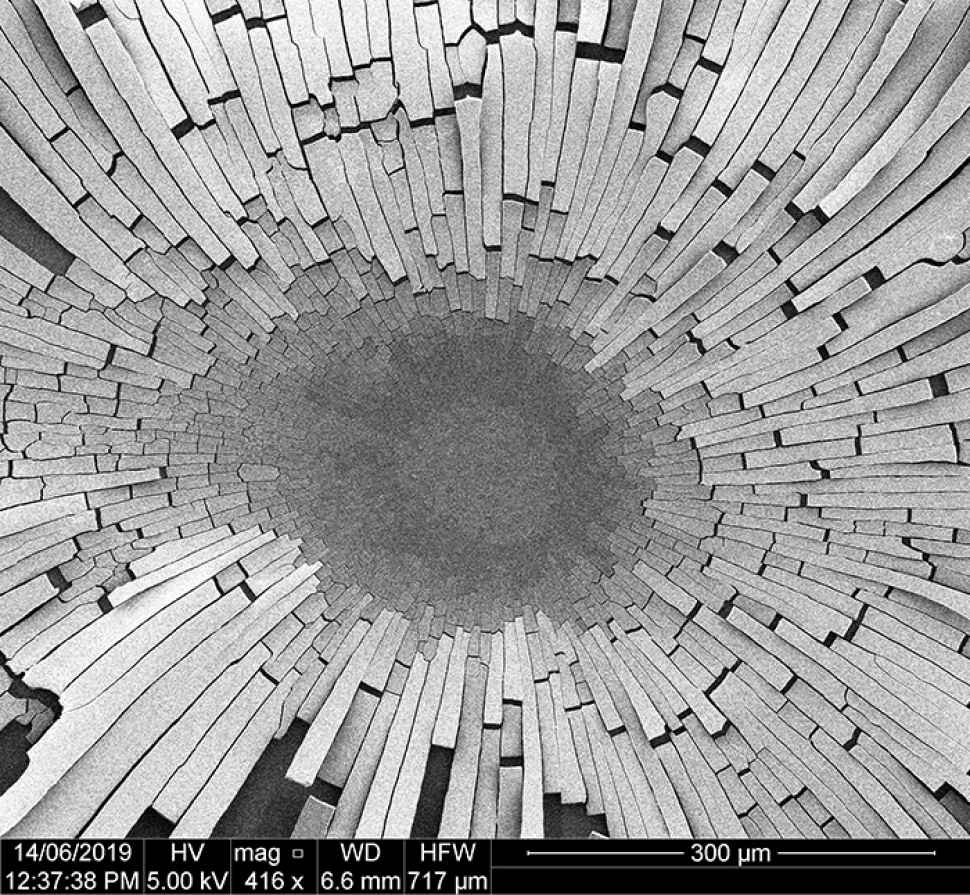
17. Split
Stefan Martino
The individual “planks” are formed from silica nanoparticles clumped together that lifted off the surface as they dried.
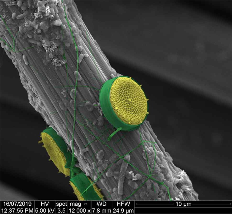
18. Seaflower – the unwanted beauty
Karuna Skipper/Sait Elmas
SEM image of diatom species on a carbon fibre. The carbon fibre is covered in extracellular polymeric substances (EPS) and bacteria (biofilm), which is the origin of marine growth (fouling). By applying only 900 millivolts vs. Ag|AgCl to the carbon cloth electrode the marine growth can be prevented.
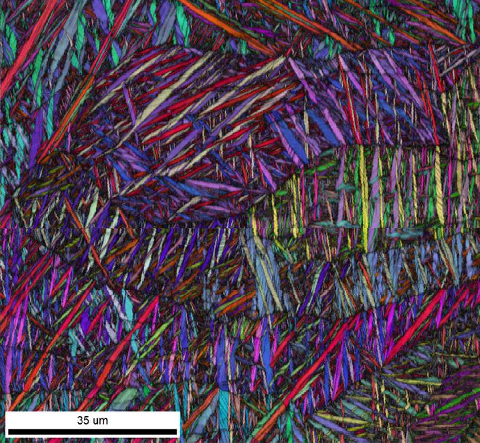
19. The colours of titanium
Aoife McFadden
We’re looking at the grain structure of titanium. The colours show us the orientation the titanium crystals are in and that tells us how strong the titanium is.
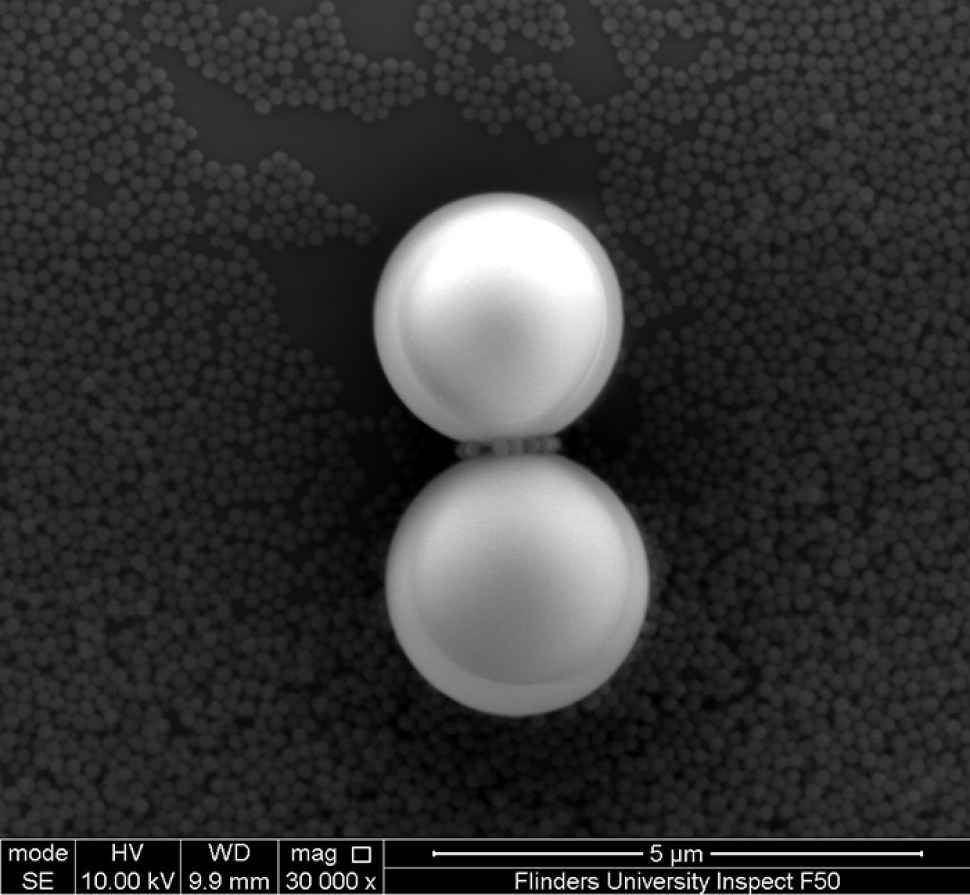
20. The (silica) spheres
Christopher Hassam
Scale model of iconic Bert Flugelman sculpture The Spheres, rendered in silica.
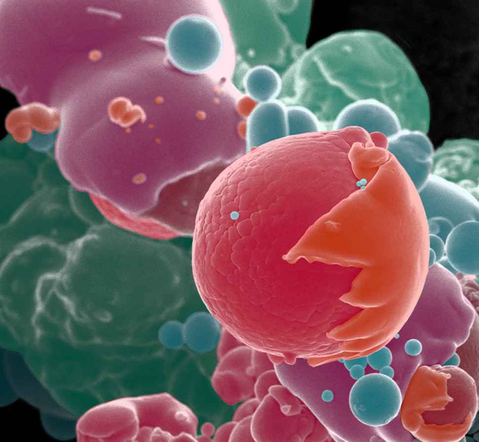
21. Who’s that Pokémon?
Dr Ruby Sims
Coloured SEM image of aluminium powder particles used as a starting material for metal LMD 3D-printing. Particles are approximately 40µm wide., FOV 100µm.
22. The dinoflagellate Alexandrium leei
Danielle Saunders
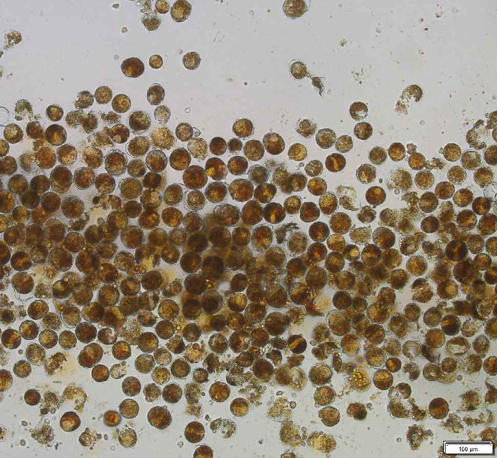
The image is of Alexandrium leei algae cells observed at 40x magnification using an IX73 Olympus Japan inverted microscope. Alexandrium leei is a non-toxic dinoflagellate, that I had the opportunity of working with whilst studying at the Institute of Oceanology Chinese Academy of Sciences (IOCAS) through the NCP program.
23. Visualization and colocalization of lipid drops in Euglena gracilis with DPAS and BODIPY
A H M Mohsinul Reza
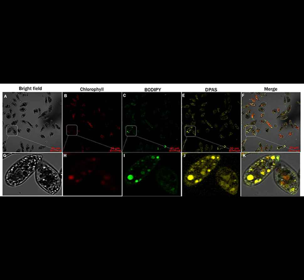
Images of Euglena gracilis cells incubated with lipid specific aggregation induced emission (AIE) nanoprobe, DPAS (C20H16N2O) and commercial lipid specific fluorescent probe, BODIPY (Bright-field image: A; Fluorescence images - Chlorophyll: B, BODIPY (Bright-field image: A; Fluorescence images - Chlorophyll: B, BODIPY: C, and DPAS: D; Merged image: E), and the enlarged regions (F, G, H, I, and J for bright-field, reduced chlorophyll, BODIPY, DPAS stained cells and merged image, respectively). Cells were cultured in modified Cramer–Myers medium (MCM), under nitrogen and calcium starved but glucose supplemented dark condition. Images were taken with Zeiss LSM 880 Airyscan confocal microscope
![]()
Sturt Rd, Bedford Park
South Australia 5042
South Australia | Northern Territory
Global | Online
CRICOS Provider: 00114A TEQSA Provider ID: PRV12097 TEQSA category: Australian University


